Case Challenge 14 – February 2020

Case Challenge 16 – July 2024

Home » Uncategorized

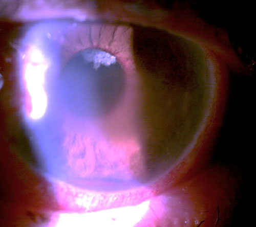
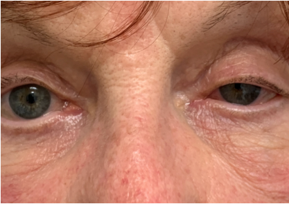
An 82 year old man presented complaining of a moderately painful left eye of approximately 2-week duration. He was both diabetic and hypertensive.  He was 20/25 OD and bare light perception OS. His intraocular pressure (IOP) was 23 mm Hg OD and 62 mm Hg OS. His left eye manifested central microcystic corneal edema, florid iris neovascularization, and hyphema. Gonioscopically, his right angle was open to at least scleral spur while his left angle was closed with peripheral anterior synechiae (PAS) for 1800 with hyphema and angle neovascularization for the remaining part of the angle. The corneal edema, circulating hyphema, and poor dilation prevent fundus examination.

Clearly this was an acutely urgent case of neovascular glaucoma (NVG). Patients with NVG typically present with a chronically red, painful eye, which often has significant vision loss.  There frequently is an antecedent history of a retinal vessel occlusion, carotid artery disease, chronic retinal detachment, or advanced diabetic retinopathy. There will be visible neovascularization of the iris (NVI) and angle (NVA).  The patient will typically have significant corneal edema, anterior segment inflammation, anterior chamber cell and flare reaction, and elevated intraocular pressure due to synechial angle closure, often exceeding 60 mm Hg.

Retinal hypoxia induces vascular endothelial growth factor (VEGF) to act upon healthy endothelial cells of viable capillaries to stimulate the formation of a fragile new plexus of vessels (neovascularization). VEGF diffuses forward to the nearest area of viable capillaries, namely the posterior iris.  Neovascularization buds off of the capillaries of the posterior iris, grows along the posterior iris, through the pupil, along the anterior surface of the iris, and then into the angle.  Once in the angle, the neovascularization, along with attendant fibrovascular support membrane will block the trabecular meshwork with progressive PAS.

Neovascular glaucoma requires prompt and aggressive therapy. This involves control of the intraocular pressure and inflammation as well as management of the retinal ischemia as well as any precipitating conditions. Upon first presentation, a strong cycloplegic such as atropine 1% BID as well as a topical steroid such as prednisolone acetate 1% or difluprednate 0.05% QID should be prescribed.  The fact that the angle may be closed with PAS does not preclude pharmacologic mydriasis from atropine. Neither does the elevated IOP disqualify steroid use. This will greatly add to patient comfort.  Aqueous suppressants in the form of beta blockers, carbonic anhydrase inhibitors, and alpha adrenergic agonists may be used in order to temporarily reduce IOP. Definitive treatment is best accomplished with pan-retinal photocoagulation (PRP) to destroy ischemic retina, minimize oxygen demand of the eye, and reduce the amount of VEGF being released. While PRP is the most definitive treatment for the neovascularization causing NVG, the advent and use of antiangiogenic drugs has proven to be a valuable adjunct. 

The patient presenting here was prescribed atropine 1% BID, prednisolone acetate 1% QID, and brinzolamide/brimonidine fixed combination TID in the left eye. Upon meeting with the retinal specialist approximately 2 weeks later, his eye pain had disappeared, his IOP was now lowered to 24 mm Hg OS, and his cornea had clear enough to allow fundus examination. There was still iris neovascularization, but the hyphema had cleared. Fundus examination of the left eye revealed combined central retinal artery and vein occlusion. He received an intravitreal injection of an anti-VEGF agent and was scheduled for PRP.

Case Challenge 15 – March 2021

Case Challenge 16 – July 2024

Home » Uncategorized
Left ptosis and miosis

A 73-year-old woman presented urgently with a sudden left ptosis. She noticed it immediately after undergoing parathyroid surgery to remove 3 adenomas the day before. At first, she and her family thought it might have been due to the surgical anesthesia, but when it didn’t resolve the next day, the surgeon directed her to the emergency room out of fear of stroke. At the ER she had a CT scan of the brain which showed no abnormalities.

When she presented for ocular consultation, she had left ptosis and miosis with pupillary dilation lag in dim light. She also mentioned that she had a headache and an ache in her left eye as well. Instillation of 0.5% apraclonidine (Iopidine) elevated her eye lid and dilated her pupil, confirming that she had a left Horner’s Syndrome.

While neck surgery was superficially sufficient to explain her new onset Horner’s syndrome, it did not readily account for her headache and eye pain. Ocular inspection showed no abnormalities to account for her ocular discomfort. Possible carotid artery dissection was discussed, but the patient was resistant to any further intervention and would not go back to the ER as she had been there earlier in the day and maintained that her imaging was normal. Review of the neuroimaging report revealed that she had a CT of the brain to rule out hemorrhagic stroke, but the area of concern in the neck was not imaged. Due to the remote possibility that she may have a carotid artery dissection, she was prescribed 81 mg aspirin daily.

Phone follow-up the next day revealed that she felt much better with her headache and ocular discomfort dissipating. She stated that she had recovered from the physical and emotional exhaustion from the surgery and ER visit. She now agreed to further assessment of her Horner’s syndrome. Magnetic resonance angiography (MRA) of the head and neck was ordered and scheduled. The results revealed that she did indeed have an acute left carotid artery dissection. Upon receiving the results, her aspirin was increased to a full 325 mg daily and she was seen by a stroke neurologist the next day. Her carotid dissection continues to heal and she therapeutically uses 0.5% Iopidine once daily in her left eye for cosmesis as it relieves her ptosis.

Horner’s syndrome is characterized by an interruption of the sympathetic nerve supply somewhere between its hypothalamic origin and the eye.  Clinical findings can include ptosis, pupillary miosis, and, in some cases, facial anhidrosis. Apraclonidine is an alpha-2 adrenergic agonist which stimulates alpha-1 receptors to a negligible degree in a normal eye. A Horner’s syndrome pupil undergoes denervation hypersensitivity with upregulation of both the number and sensitivity of available receptors. When a very weak alpha-1 adrenergic agonist is applied, the hypersensitive pupil dilates while the normal pupil has no effect.   Pupil dilation in suspected Horner’s syndrome is considered diagnostic. Additionally, ptosis also markedly improves with this medication.

In patients with new onset Horner’s syndrome concurrent with recent ipsilateral neck trauma, neck and face pain, ipsilateral transient monocular visual loss, or contralateral transient weakness or numbness, acute cervical carotid dissection must be immediately suspected. Carotid artery dissection is a linear tear in the vessel wall that allows for thrombus formation and possible subsequent embolization. In this case, there is a substantial risk of hemispheric stroke within the first two weeks of onset.  Cervical carotid artery dissection is a relatively common cause of acute onset Horner’s syndrome. As the sympathetic fibers travel with the carotid artery within the neck, damage to the vessel can result in Horner’s syndrome. New onset Horner’s syndrome with head, neck, eye or face pain or after recent neck trauma must be emergently evaluated for carotid artery dissection. Patients will need stroke prophylaxis while the vessel wall heals and embolic risk dissipates.

Case Challenge 16 – July 2024

Case Challenge 16 – July 2024

Home » Uncategorized

A 46-year-old woman presents for new onset vision loss in her right eye for the past month. She has no pain or diplopia and feels that her vision has been stable since. Her medical history is unremarkable and felt that she might have had COVID one week before vision loss, but did not test for the virus. Her best corrected visual acuity is 20/60 OD and 20/20 OS with a relative afferent pupil defect OD. Her dilated exam reveals normal optic discs with cupping at 0.6/0.6 OD, OS. Optical coherence tomography (OCT) reveals a normal retinal nerve fiber layer and ganglion cell complex OU. Threshold perimetry is normal in her left eye and there is an inferior arcuate defect in her right eye.

While it is tempting to initially diagnose this as retrobulbar optic neuritis, possibly associated with multiple sclerosis, it would be a superficial assessment. The lack of pain is concerning as the vast majority of patients with retrobulbar demyelinating optic neuritis complain of pain. A painless loss of inferior visual field can often occur from non-arteritic anterior ischemic optic neuropathy, but the normal disc appearance and lack of a disc-at-risk mitigated against this diagnosis.

Upon further discussion, she acknowledged that she felt that she was having trouble with her right contact lens for several months. She acknowledged that her vision loss may have been present for longer than the past month and that she may had just become aware of it. Contrast enhanced magnetic resonance imaging of the brain, orbits, and chiasm revealed a planum sphenoidale meningioma compressing the prechiasmatic portion of the right optic nerve. Subsequent craniotomy with tumor removal was successful, with her visual acuity improving to 20/15 and her visual field loss totally resolved. This was anticipated as her OCT findings were normal, indicating that structurally her visual system was intact though she had functional loss on visual fields.